At least 30 to 40 percent of bone mineral must be lost before bone loss is detectable on a routine X-ray, and that is too late for an effective prevention of fracture risk. Specialized tests have been developed to detect small amounts of bone mineral loss in those people at risk. The most widely available and preferred technique is Dual-Energy X-ray Absorptiometry or DXA.
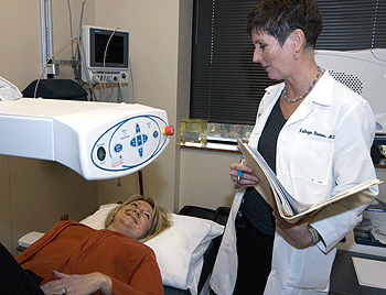
During the DXA examination, the patient lies on a padded table while a “scanning post” passes back and forth over the site of the skeleton being measured. No injections or dyes are used. A simple and fast procedure, the DXA is a comfortable diagnostic test that measures bone mass in the spine, hip and forearm.
During a five- to seven-minute period of time, an X-ray beam records the amount of calcium in the skeleton. The DXA machine’s computer, then, calculates the amount of bone mineral and compares it to values considered normal for persons of the same gender as the patient.

The amount of X-ray received is minimal — only one-sixth that of a chest X-ray or what a person might be exposed to from the altitude while flying from New York to Los Angeles. Even though the X-ray exposure is minimal, DXA of the hip and spine is not performed on pregnant women as a matter of institutional health policy. Bone mineral density measurements of the forearm may be used in pregnant women.
A trained Bone Density Technologist certified by the International Society for Clinical Densitometry (ISCD) performs the DXA scan. He or she will explain the procedure to you. The specialist physicians at the Bone Health Program, most of whom are ISCD Certified Clinical Densitometrists, will interpret the results and discuss it with you at the visit, in most cases immediately after the test. The physician will also submit a report including detailed measurement data to your physician within a few days.
The DXA provides a safe, inexpensive and accurate means of measuring bone mineral density. Similar to a baseline mammogram, the initial DXA provides a baseline measurement or point of comparison for future measurements.
Repeated measurements at specified intervals determine an accurate rate at which bone is being lost and provide a means to monitor the effectiveness of treatment. In order to most accurately estimate changes in bone density between scans, it is strongly recommended that you obtain your bone density test at the same facility and on the same machine. This is because DXA tests performed at different centers cannot be directly compared. To ensure the best accuracy in follow-up tests, all the DXA machines
operated by the bone health program are cross-calibrated and subjected to frequent rigorous precision testing.
The DXA measurement helps physicians assess the likelihood of developing fractures that may occur during normal living activities, or after minimal trauma. For example, more than 30 percent loss of bone mass in the hip leads to a significant increase in the fracture rate.
Under the aegis of the World Health Organization, a tool has been developed, the FRAX calculator that aids in estimating fracture risk based on bone density but also taking into account other factors that are known to increase fracture risk, such as family history of osteoporosis, having had a previous low-trauma fracture, taking medications that negatively affect bone strength (i.e. corticosteroids for rheumatoid arthritis, lupus, or asthma), smoking, and others. While this tool is freely available online, we strongly encourage you to consult with your specialist physician or health care provider to devise the most appropriate approach to improve and maintain your bone health.
Making an appointment is easy, please call 314-454-7775. Appointments can be scheduled usually within two weeks. During the first phone call, you will be asked the name of your referring physician, and other basic information about yourself. If you need a referral from your insurance company, please obtain authorization before your appointment.
You do not have to prepare anything special for the DXA. Keep to your normal diet and daily routine. The whole DXA takes 20 minutes from start to finish, including filling out any of the other required forms. To help with the paperwork, you can download and fill out a simple Patient Questionnaire (pdf) and Medication Record (pdf).
To make the examination easier, we recommend that patients wear loose fitting comfortable clothing, such as sweatshirts and sweat pants with no zippers, buttons, or safety pins, or other metal fasteners. Patients may be asked to change into a hospital gown. Please do not take calcium or OTC potassium supplements. If you are taking prescription potassium, please contact your prescribing physician if you can skip your dose(s) 24 hours prior to your test.
Our division has a powerful tool to enhance the assessment of skeletal health in our patients – Trabecular Bone Scores (TBS). This advanced imaging software offers valuable insights into bone microarchitecture, making it an essential addition to our repertoire of diagnostic technologies.
TBS is an innovative textural index that provides an indirect indicator of bone microarchitecture. This cutting-edge software evaluates pixel gray-level variations in the lumbar spine DXA (dual-energy X-ray absorptiometry) image. By analyzing the texture of the bone, TBS can assess the strength and density of trabecular bone – the inner, spongy part of bones responsible for supporting bone mass.
One of the remarkable aspects of TBS is its applicability to a broad range of patients. It is appropriate for all patients whose spines are evaluable and have at least two vertebrae available for analysis. However, there are certain cases where spine measurements and TBS may be excluded, such as in the presence of hardware, scoliosis, or extreme degeneration. In these situations, alternative methods may be employed to assess bone health.
Extensive cross-sectional and longitudinal studies have demonstrated that TBS can independently predict fragility fractures. This ability to assess bone microarchitecture provides valuable information that complements traditional bone mineral density (BMD) measurements. TBS offers a more comprehensive view of skeletal health, allowing for early identification of individuals at risk of fractures, thus facilitating proactive preventive measures.
Beyond predicting fractures, TBS can be used to adjust FRAX probabilities of fracture. FRAX (Fracture Risk Assessment Tool) is a widely utilized tool for assessing the ten-year probability of a major osteoporotic fracture in an individual. By incorporating TBS data, the accuracy of FRAX calculations can be further refined, leading to more informed decisions regarding patient management.
TBS proves to be a valuable asset in monitoring the response to pharmacologic treatment for osteoporosis. As patients undergo treatment, changes in bone microarchitecture can be tracked over time, providing insights into the effectiveness of prescribed therapies. This ability to gauge treatment efficacy helps tailor interventions to individual patient needs, leading to improved outcomes.
Usefulness in Secondary Osteoporosis
Certain patient populations with secondary osteoporosis may benefit greatly from TBS. Secondary osteoporosis refers to bone loss resulting from underlying conditions or medications. By assessing bone microarchitecture, TBS can offer additional information in these complex cases, aiding in more precise diagnoses and treatment plans.
The acquisition of Trabecular Bone Scores (TBS) represents a significant advancement in our ability to assess skeletal health. By evaluating bone microarchitecture, TBS complements traditional BMD measurements, enabling more accurate prediction of fragility fractures and facilitating individualized treatment plans. Moreover, its utility in monitoring treatment response and its application in cases of secondary osteoporosis further underscore the value of TBS in patient care. We look forward to leveraging this powerful imaging software to enhance the overall well-being and quality of life of our patients.
Washington University School of Medicine does not endorse or guarantee the accuracy of information contained on websites on non-affiliated external sources. Read the School of Medicine’s Policy on Links to Third-Party Websites to learn more.